


Actin Structure, Dynamics and Regulation

Uncovering the molecular mechanisms underlying actin regulation and force generation in Apicomplexan parasites
Force generation and motility are central to the life cycle of Apicomplexan parasites, a group of protozoans responsible for major health and economic problems worldwide. All these parasites rely on specialized molecular machinery, including divergent actomyosin complexes, to move, invade host cells and complete their life cycle. Understanding how this machinery works in Plasmodium, the causative agent of malaria, could allow to identify novel antimalarial targets.
Actin is a globular protein that can polymerize into filaments that work with myosin motors to generate force. Apicomplexan parasites have evolved an unusual actin cytoskeleton with properties that differ substantially from those found in other eukaryotes. Apicomplexan actin is highly dynamic and does not form stable filaments in vitro. Many questions remain unanswered, for instance : “How do parasites control actin assembly and disassembly in vivo?” “How are actin filaments organized in space and time to produce force exactly when and where it is needed?
Plasmodium falciparum expresses two divergent actins, PfAct1 and PfAct2, which contribute to different stages of the parasite life cycle. The parasite relies on a limited repertoire of actin-binding proteins (PfABPs) to regulate its actin cytoskeleton. Most, if not all, PfABPs are essential for the survival of the parasite and differ markedly from their counterparts in other eukaryotes. How these proteins interact with actin, regulate filament dynamics and coordinate force generation remains poorly understood.
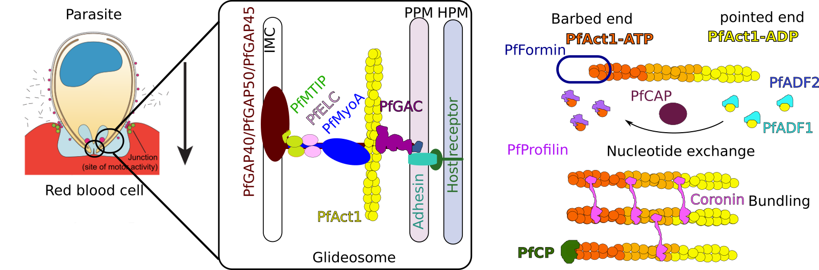
Regulation of the actin cytoskeleton in Plasmodium falciparum. From left to right: schematic representation of a merozoite invading a red blood cell. A key element of invasion is a macromolecular complex called the glideosome, located between the inner membrane complex (IMC) and the parasite plasma membrane (PPM). The glideosome is organized around the unconventional myosin A (PfMyoA) and short PfAct1 filaments. PfAct1 filaments are bound to PfGAC. On the right, a representation of PfABPs expressed by P. falciparum and their putative roles in PfAct1 regulation.
In ACTOMOVE, we aim to uncover the molecular mechanisms that govern the regulation of the Apicomplexan actin cytoskeleton. We combine structural biology approaches, such as X-ray crystallography and cryo-electron microscopy, with molecular dynamics simulations and in vitro biochemical and biophysical assays. By bridging these complementary approaches together, we investigate how specific Apicomplexan ABPs interact with actin and control its assembly, organization and dynamics.
Our research is part of a broader effort to connect molecular mechanisms with biological function. Through collaborations with an international network of experts, we seek to link our molecular insights to advanced experimental approaches that investigate actin regulation in vitro and in vivo. By revealing how Apicomplexan actin cytoskeleton is regulated, ACTOMOVE aims to advance our understanding of parasite biology and identify potential pharmaceutical targets that could lead to new strategies to fight malaria and other Apicomplexan-associated diseases.
team

Group Leader Researcher
External funding

ANR JCJC