The Electron Microscopy facility of Imagerie-Gif is dedicated to Cell Biology.
It includes all the equipment required to prepare samples, from nanoparticles to tissues, with a large variety of techniques, from routine methodology (chemical fixation, high-pressure freezing, ultramicrotomie, immunolabeling, cryo-EM) to advanced modalities (CLEM, tomographie, 3D reconctruction)
More than providing an up-to-date equipment to prepare and image sample at the nanoscale resolution, Imagerie-Gif promotes the training of academic and private researchers via national workshops and on-demand trainings.
R&D Axes
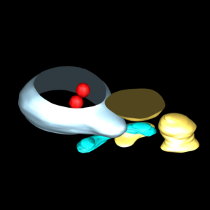
3D recontruction
FIB-SEM
FIB-SEM allows serial section acquisition of biological sample after conventional resin embedding. The serial images are then aligned and processed to obtain a 3D reconstruction of large volumes at nanoscale resolution. FIB-SEM can be correlated with fluorescence microscopy to optimise the localisation of specific events.
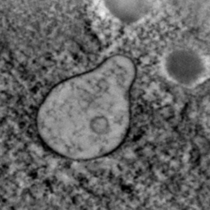
Tomography
Electron tomography is technique for obtaining detailed 3D structures of sub-cellular specimensThe electron beam is passed through the sample at incremental degrees of rotation. This information is collected and used to assemble a three-dimensional image of the target. For biological applications, the typical resolution of ET systems are in the 5-10nm range.
Conventional tomography is usually suitable for 150nm-thick sections. The thickness can be increased if the tomogram is acquired with the STEM detector.
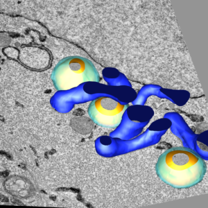
Serial sections
Serial sectioning is a method to generate high resolution three-dimensional images from small samples. The technique is widely applicable for any biological samples embedded in resin. Serial ultrathin sections are mounted on a grid, and images of these sections are obtained by TEM. The combination of all images obtained on all sections provides 3D-structures of the sample.
chemical mapping
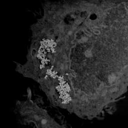
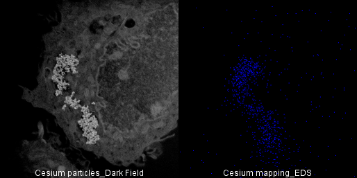
Chemical mapping
Energy-dispersive X-ray spectroscopy (EDXS) is an analytical technique used for the chemical characterization of a sample. It relies on an interaction of some source of X-ray excitation and a sample.
When exposed to the electron beam, the sample emit an electromagnetic emission spectrum in which the peak positions are specific to its elemental composition. Combining EDXS with STEM imaging allows chemical mapping of the sample.
correlative microscopy
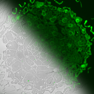
Correlative Light and Electron Microsopy
Correlative light-electron microscopy (CLEM) is the combination of an optical microscope – usually a fluorescence microscope- with an electron microscope. Suitable protocols allow the preservation of fluorescence and of sample orientation between fluorescence image and electron micrograph. Overlay of the two images is thus performed as a result of the integration of 2 microscopes. Imagerie-Gif benefits from the expertise of both optical and electron microscopy on the same site and performs CLEM experiments adapted to each problematic.