Fluorescent biosensors are molecules and proteins that are sensitive to one or multiple environmental parameters. This characteristic will directly influence their capacity to emit fluorescence, which will allow us to quantify the variations of this parameter. Many types of biosensors are today well described, allowing the intra cellular investigation of a broad variety of molecules and phenomena: calcium concentration, reactive oxygen species production, pH variations, membrane tension…
Multiple approaches are developed on the facility to assist the users at different steps of biosensors experiments: – choice of the biosensor (inorganic or genetically encoded, ratiometric or lifetime-based) – setting up an optimal image acquisition on the microscope – generation of a calibration curve to reach absolute measurements – design of data analysis pipelines
Fluo4_full.gif
Detection of cytosolic calcium variations in HeLa cells using the FLUO-4 dye.
Ratio_pHP_Bacteria_Web.gif
Detection of pH variations in Sinorizobium meliloti expressing the genetically encoded biosensor pHP
Super-resolution microscopy
Super-resolution microscopy is a family of technics (PALM, SIM, STED…) allowing to break the diffraction barrier that limits the resolution of conventional microscopes around 250nm. Although these techniques are very powerful (XY resolution is increased up to 10 times), they are nevertheless very complex to implement. The facility offers the possibility to perform PALM/dSTORM, Lattice SIM and Live-SR depending of the question and the limitations of the sample. Engineers have developed methods and tools to assist the users in the sample preparation, image acquisition, image reconstruction and quantification.
Live-SR is a module implemented on a spinning disk, allowing to increase the XY resolution up to 150 nm without any limitation concerning the fluorescent tags used, the frame rate or the sample preparation.
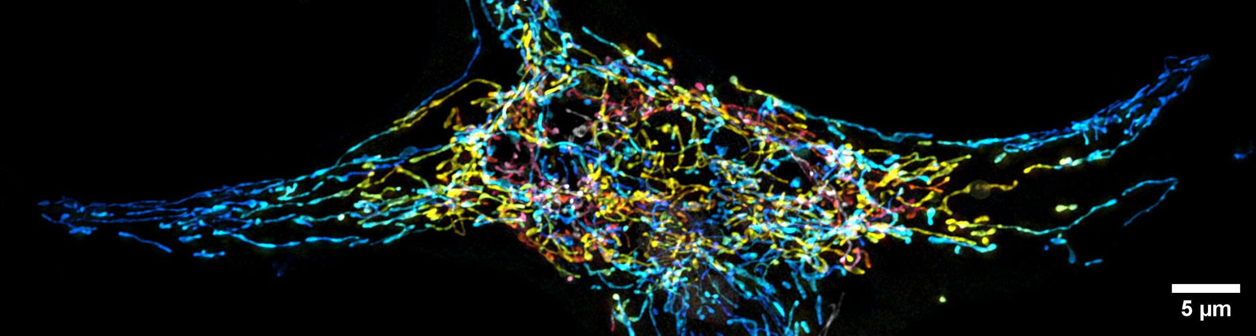
Endoplasmic reticulum labelled with GFP imaged in an Arabidopsis thaliana root epidermis.
Lattice SIM is also a versatile technique allowing to reach resolution up to 100 nm with any fluorescent probes on both live and fixed samples. Due to the lattice illumination pattern, this new generation of SIM is more efficient to generate 3D super resolution images and more suited to go deeper in a sample.
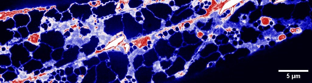
Mitochondrial network of a HeLa cell.
PALM & dSTORM is one of the most powerful microscopy technique with an achievable resolution around 40 nm in XY. It is the method of choice to reach the molecular level within a cell to unveil nanometric architecture. Nevertheless, this approach requires a specific sample preparation and is limited to the use of certain fluorescent probes. Furthermore, as several minutes are needed to generate PALM or dSTORM images, this technique is not suited for the study of dynamic processes.
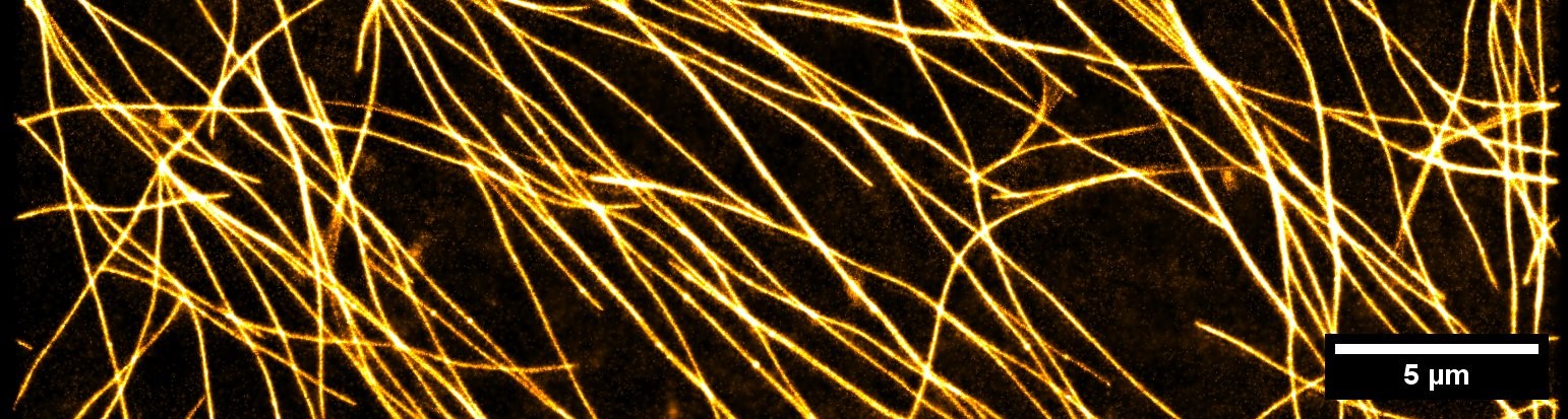
Microtubules labelled with mEOS, imaged in PALM in an etiolated Arabidopsis thaliana hypocotyl.