Function and architecture
of macromolecular assemblies

Research Projects

Molecular mechanisms of the genome replication initiation and restart in bacteria

Principal Investigators: Sophie Quevillon-Cheruel, Stéphanie Marsin and Hélène Walbott

Engineers involved: Inès Li de la Sierra-Gallay and Magali Aumont-Nicaise

PhD student: Claire Cargemel
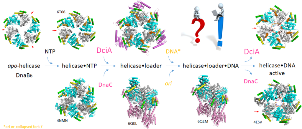
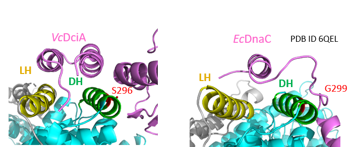
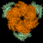
In all living organisms, replicative helicases are essential proteins that unwind double stranded DNA in front of replication forks. The loading of the helicases depends on accessory proteins such as DnaC and DnaI which are two well characterized loaders in bacteria. However, in most bacteria these loaders are replaced by the unrelated and essential protein DciA, recently discovered by Jean-Luc Ferat (Genomes Department I2BC). The goal of our work is to understand the molecular mechanisms that lead to the loading of dciA-replicative helicases on DNA, and to describe at the molecular level the induced structural rearrangements. The crystal structures of DnaB/DciA obtained so far combined with biochemical and biophysical data demonstrated the functional similarities between the dnaC/I– and the dciA-helicases, but also highlighted differences in their biochemical activities (loading, translocation, ATPase activity, 3D structures at different steps…).
The structural data revealed unexpected similarities for the mode of interaction between the helicase and DnaC or DciA, despite the total lack of structural homology between these two proteins.
This discovery opens opportunities for the development of mimicking peptides that prevent the interaction between the two proteins. These peptides could prevent the replication of the genome and hence eradicate pathogenic bacteria.
Publications

Marsin S, Adam Y, Cargemel C, Andreani J, Baconnais S, Legrand P, Li de la Sierra-Gallay I, Humbert A, Aumont-Nicaise M, Velours C, Ochsenbein F, Durand D, Le Cam E, Walbott H, Possoz C, Quevillon-Cheruel S*, Ferat JL*.

Study of the DnaB:DciA interplay reveals insights into the primary mode of loading of the bacterial replicative helicase.

(2021) Nucleic Acids Res. 49(11):6569-6586.

https://pubmed.ncbi.nlm.nih.gov/34107018/


Chan-Yao-Chong M, Marsin S, Quevillon-Cheruel S, Durand D, Ha-Duong T.

Structural ensemble and biological activity of DciA intrinsically disordered region.

(2020) J Struct Biol. 212(1):107573.

https://pubmed.ncbi.nlm.nih.gov/32679070/


Brezellec P, Vallet-Gely I, Possoz C, Quevillon-Cheruel S, and Ferat J-L

DciA is an ancestral replicative helicase operator essential to bacterial replication initiation.

(2016) Nature Communications. 7:13271.

https://pubmed.ncbi.nlm.nih.gov/27830752/

Molecular mechanism of cell-cell communication in Gram-positive bacteria

Principal Investigator: Sylvie Nessler

Ingeneers involved: Noureddine Lazar, Inès Li de la Sierra-Gallay

Former students involved: Rosa Granha, Samira Zouhir, Antoine Talagas, Jordhan Thuillier
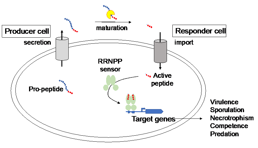
The search for new antimicrobials has highlighted the therapeutic potential of inhibitors of bacterial cell-cell communication systems designated as quorum-sensing (QS) systems. Through secretion of diffusible signal biomolecules called pheromones, QS coordinates the behavior of a whole community of bacteria in response to environmental conditions. In Gram-positive bacteria, re-internalized peptide pheromones directly control the activity of cytoplasmic transcriptional regulators.
These quorum sensors control important bacterial processes such as virulence, biofilm formation, necrotrophism and sporulation. We structurally and functionally characterized several Gram-positive quorum-sensors and showed that they form a conserved family called RRNPP, characterized by a TPR-type peptide-binding domain and a HTH-type DNA-binding domain.
Using structural and functional analyses, we showed that, despite divergent protein and peptide sequences, the binding mode of the peptide is conserved among RRNPPs. The activation mechanism is however specific to each member of the family.
Our objective is now to exploit our knowledge on RRNPP-type QS regulators to design inhibitors based on synthetic peptide derivatives.
Publications

Molecular dissection of pheromone selectivity in the competence signaling system ComRS of streptococci. 

Ledesma-Garcia et al. (2020) Proc Natl Acad Sci USA. 117:7745-7754

Subtle selectivity in a pheromone sensor triumvirate desynchronizes competence and predation in a human gut commensal. 

Mignolet et al. (2019) eLife 8: e47139

Structural Insights into Streptococcal Competence Regulation by the Cell-to-Cell Communication System ComRS.  

Talagas et al. (2016) PLoS Pathog. 12:e1005980 

Pheromone Recognition and Selectivity by ComR Proteins among Streptococcus Species.

Shanker et al. (2016) PLoS Pathog. 12:e1005979

NprR, a moonlighting quorum sensor shifting from a phosphatase activity to a transcriptional activator.

Perchat et al. (2016) Microb Cell. 3:573-575

How Quorum Sensing Connects Sporulation to Necrotrophism in Bacillus thuringiensis

Perchat et al. (2016) PLoS Pathog. 12:e1005779

Peptide-binding dependent conformational changes regulate the transcriptional activity of the quorum-sensor NprR.

Zouhir et al. (2013) Nucleic Acids Res. 41:7920-33

Activity of the Bacillus thuringiensis NprR-NprX cell-cell communication 1 system is coordinated to the physiological stage through a complex transcriptional régulation

Dubois et al. (2013) Mol Microbiol. 88: 48-63

Structural basis for the activation mechanism of the PlcR virulence regulator by the quorum-sensing signal peptide PapR

Grenha et al. (2013) Proc Natl Acad Sci USA. 110: 1047-52

A cell-cell communication system regulates protease production during sporulation in bacteria of the Bacillus cereus group

Perchat et al. (2011) Mol Microbiol. 82: 619-633

The universal t6A modification pathways of ANN-accepting tRNAs in the three domains of life.

Herman van Tilbeurgh : principal investigator

Dominique Liger : associate professor

Bruno Collinet : associate professor

Sylvie Auxilien : researcher

Noureddine Lazar : researcher

Charles Cirio : PhD student

Raoudha Dammak : technician
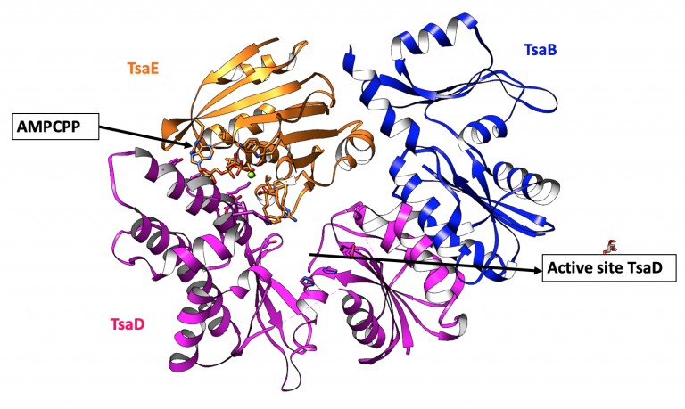
The N6-threonylcarbamoyl adenosine modification (t6A) is one of the few universally conserved tRNA modifications. It adds a threonylcarbamoyl moiety on position 37 of tRNAs that read ANN codons. In vivo, t6A is important for start codon recognition and reading frame maintenance. The t6A system mobilizes partially different sets of proteins in bacteria on the one hand and archaea/eukaryotes on the other. In E. coli, apart from TsaD (=Kae1) and TsaC (=Sua5) two more proteins are essential for the t6A modification : TsaE and TsaB. Our recently determined 3D structure of the ternary TsaB/D/E complex partly explained how this sophisticated machinery might work. The main objective of this project is to further unravel the molecular/mechanistic aspects of the bacterial t6A system. The t6A system is essential for most bacteria and therefore offers opportunities for antibiotics development. We are collaborating with chemists to develop compounds that specifically block the t6A activity in bacteria.
Structure of the bacterial Tm TsaB/D/E complex
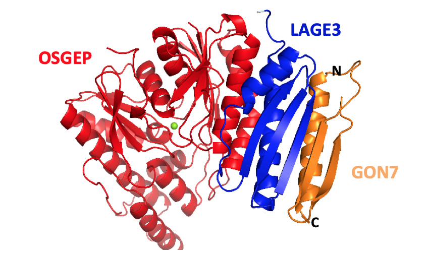
In archaea and eukaryotes, this modification is catalyzed by Kae,1 which is the homolog of bacterial TsaD. Kae1 is part of a stable multiprotein complex KEOPS, together with four other proteins : Gon7, Bud32, Pcc1 and Cgi121. Particular mutations in the five human genes coding for all these subunits (Gon7, TP53K, OSGEP, LAGE3 and TPRKB) are responsible for the Galloway-Mowat syndrome (GAMOS), a clinically heterogeneous autosomal recessive disease characterized by early-onset steroid-resistant nephrotic syndrome and microcephaly. Our structure of a partial human KEOPS demonstrated that these mutations are spread out over the complex. Using biochemistry, biophysics and structural biology (crystallography and cryo-EM) we endeavor to understand the molecular underpinning of this serious disease and in particular the role of the 4 other subunits of the KEOPS complex. Another main objective of this project is therefore to unravel the molecular/mechanistic aspects of the t6A system in eukaryotes and archaea.
The human KEOPS subcomplex
Publications

Structure of a reaction intermediate mimic in t6A biosynthesis bound in the active site of the TsaBD heterodimer from Escherichia coli.

Kopina et al. (2021) Nucleic Acids Res. 49:2141–2160

Defects in t6A tRNA modification due to GON7 and YRDC mutations lead to Galloway-Mowat syndrome.

Arrondel et al. (2019) Nat Commun. 10:3967.

Mutations in KEOPS-complex genes cause nephrotic syndrome with primary microcephaly.

Braun et al. (2017) Nat Genet. 49:1529-1538.

The structure of the TsaB/TsaD/TsaE complex reveals an unexpected mechanism for the bacterial t6A tRNA-modification.

Missoury et al. (2018) Nucleic Acids Res. 46:5850-5860

Crystal structures of the Gon7/Pcc1 and Bud32/Cgi121 complexes provide a model for the complete yeast KEOPS complex.

Zhang et al. (2015) Nucleic Acids Res. 43: 3358–3372.

The ATP-mediated formation of the YgjD-YeaZ-YjeE complex is required for the biosynthesis of tRNA t6A in Escherichia coli.

Zhang et al. (2015) Nucleic Acids Res. 43:1804-1817.

In vitro biosynthesis of a universal t6A tRNA modification in Archaea and Eukarya.

Perrochia et al. (2013) Nucleic Acids Res. 41:1953-1964.

Gcn4 misregulation reveals a direct role for the evolutionary conserved EKC/KEOPS in the t6A modification of tRNAs.

Daugeron et al. (2011) Nucleic Acids Res. 39:6148-6160.

Structure of the archaeal Kae1/Bud32 fusion protein MJ1130 : a model for the eukaryotic EKC/KEOPS subcomplex.

Hecker et al. (2008) EMBO J. 27:2340-2351.

An archaeal orthologue of the universal protein Kae1 is an iron metalloprotein which exhibits atypical DNAbinding properties and apurinic-endonuclease activity in vitro.

Hecker et al. (2007) Nucleic Acids Res. 35:6042-6051.

Maturation and degradation of tRNA by the yeast RNaseZ (Trz1)

Team members involved: Ines Li de la Sierra-Gallay, Noureddine Lazar & Herman van Tilbeurgh
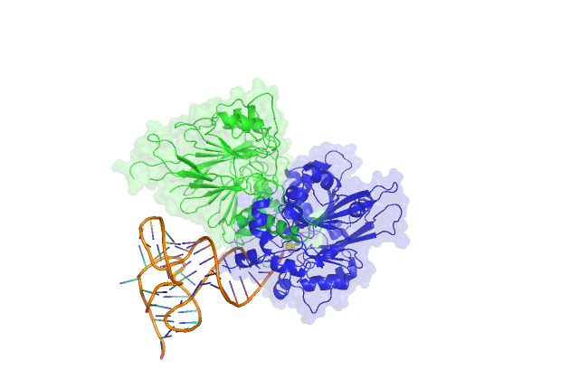
RnaseZ is an essential and highly conserved protein, present in bacteria, archaea and eukaryotes. The enzyme is a 3’ endonuclease that cleaves precursor tRNAs that are missing the CCA motif next to the discriminating base. Two RnaseZ types are present in eukaryotes: a short and a long form (RNaseZ(L)). The long form is composed of two domains: a C-terminal domain that contains the catalytic activity and an N-terminal domain that modulates the specificity of the enzyme. Our crystal structure of Trz1, the yeast long form RnaseZ, has shown that the two domains have a common fold, very similar to that of the short form RnaseZ (1-3).
Trz1 from yeast was shown to form a tight complex with two other enzymes, Nuc1 and YMR099c. Nuc1 is an endonuclease and an ortholog of the human ENDOG, involved in the recombination of mitochondrial DNA and in cellular apoptosis. YMR099cis hexose-phosphate mutarotase, whose structure was also determined in our team (4). Despite the fact that the complex very likely exists in cellulo, its biological function remains a mystery. Our team endeavors to obtain the cryo-EM structure of this complex (5). Structural data will be exploited to unravel the function of this very tight complex using biochemical and cellular approaches.
3D-model of Trz1 in complex with its tRNA substrate (the N- and C-termial domain are in green and blue respectively).
Publications
Avirulence effectors of the phytopathogenic fungus Leptosphaeria maculans: structural and functional characterization

Team members involved: Karine Blondeau, Noureddine Lazar, Ines Li de la Sierra-Gallay & Herman van Tilbeurgh
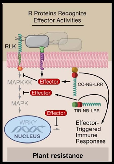
The Leptosphaeria maculans ascomycete fungus is the causal agent of one of the most devastating colza.

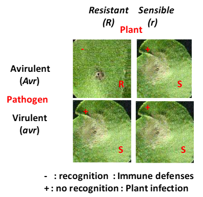
During infection, the Leptosphaeria maculans pathogen secretes an arsenal of small proteins (SSPs) or effectors, capable of modulating the response or immunity of the plant and of facilitating fungus infection. Some of these effectors, called avirulence effectors (AVR); are involved in a specific response of the plant against the infection (ETI for effector triggered immunity). The AVR proteins are targeted by plant resistance proteins (R). The avirulence proteins are usually small cysteine rich proteins that do not display sequence homologies with other proteins and hence their biochemical function is therefore difficult to predict. Also, their interactions (direct or indirect) with plant target proteins are poorly understood. The 3D structure determination of the avirulence effectors has proven to be instrumental for the comprehension of their biological function. We combine structural and biochemical approaches to study the avirulence effector AvrLm4-7 (Blondeau, Blaise et al., 2015). A few more effector protein structures have been determined since, using procaryotic or eukaryotic expression systems. These structures were very informative on the mutual functional relationships of these effectors. A new project has been initiated to study the interactions of these effectors with their resistance and or target plant proteins.
Publications

Methodologies

Biochemistry and Molecular Biology

  • Cloning, Site-directed mutagenesis, CRIPSR-Cas9 (yeast)
  • Production of recombinant proteins (expression in bacterial and eukaryotic hosts, fermentation)
  • Protein and protein complex purification
  • Enzymology studies
  • Interaction measurement (SEC-MALS, ITC, DSC, SPR)

Structural Biology

  • Crystallogenesis
  • X-ray crystallography (Regular access to SOLEIL and ESRF synchrotron facilities)
  • Small-angle X-ray scattering (SAXS)
  • Cryo-electron microscopy

Past members of the team

Joel Janin

Marc Graille

Nicolas Leulliot

Cécile Mérigoux
Anne Poupon

Ilham Aboulfath

Sophia Missouri

Gregory Deicsics

Maud Chan-Yao-Chong

Louisa Celma

Jordhan Thuillier

Adeline Pichard Kostuch

Wenhua Zhang

Antoine Talagas

Miao Ma

Dyana Sanchez

Johnny Lisboa

Mounira Tiouajni

Zaineb Fourati

Juliette Létocart

Julien Henri

Marion Boudes

Julie Bernauer

Karen Coeytaux

Thomas Bourquard

Jenny Keller

Lionel Tresaugues

Mark Brooks

Andrea Cavagnino

Lionel Cladière

Philippe Savarin

Sabrina Haquin

Eric Oeuillet

Isabelle Krimm 

Zhan-Cao Zhou

Isabelle Sorel

François Lecointe

Vincent Bondet

Kerstin Koch

Philippe Meyer

Mouna Raji

Salim Bouhazam

Liza Ammar-Khodja

Julien Vercruyssen

Samia Ben Rejeb

Samuel Le Cam

Norik Lexa-Sapart

Toufic El Arnaout

Amina Serour

Mickel Bremang

Pierre-Yves Boyer

Emilie Zelie (CDD AI)

Théophile Marsolier (L2 DEUST)

Marion Dos Santos Malhao (CDD AI)

Stéphane Plancqueel (CDD AI)

Isslam Bouazzaoui (M1)

Désiré MAHDI MOUSSA (CDD AI)

Halil Bounoua (L2 bio+)

Théo Lemoigne (CDD AI)

Joseph Bareille

Maxime Chaillet

Benjamin Dray

Céline Pinaglia (CDD AI)

Nathalie Ulryck (CDD AI)

Jerome Cicolari (CDD AI)

Hong Nhung vu (L2 DEUST)

Audrey Labarde (CDD AI)

Sami Kolli (L2 bio+)

Maelys LEJEUNE (L2 DEUST)

Emmanuel Daguet (L2 DEUST)

Julie Paulinyce (L2 DEUST)

Seiki  ACHIEDO (L2 DEUST) 

Lila Delbos (CDD AI)

Anthony Doizy (CDD AI)

Raja Dey (CDD AI)

Maxime Chaillet (CDD IE)

Charlotte Saint André (CDD AI)
Bruno  Faivre (CDD IE)
Chloé BERTHE (M1)
Maylis Lejeune (DEUST stage par alternance)
Seiki Achiedo (DEUST et L3 pro apprentissage)

Scroll to Top