Integrity of genome and polarity in bacteria
Topics
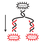
Worldwide dissemination of multidrug resistance (MDR) within bacterial populations is a major issue in public health. The main driving force for such MDR dissemination is the horizontal gene transfer (HGT) of mobile genetic elements (MGE), mainly conjugative plasmids carrying MDR genes. Conjugative plasmids encode genes necessary and sufficient for their direct cell-to-cell transfer (conjugation) to propagate within bacterial populations but also for their faithful maintenance (segregation) which ensures efficient vertical gene transfer to daughter cells at cell division. Therefore, a cell newly acquiring a conjugative plasmid turns into a stable new donor and contribute to exponential MDR wide-spreading.
Our lab is interested in factors and mechanisms involved in the prevalence of MDR conjugative plasmid, encoded in plasmid, donor chromosome, and recipient genome.
Our recent studies highlight important molecular reactions happening in the recipient cell after the entry of single-stranded plasmid DNA, which are critical for successful establishment of the plasmid in the new host (Fomenkov et al., 2020; Couturier et al., 2023; Shen et al., 2023).
Selected publications:
Yamaichi, Y., Chao, M.C., Sasabe, J., Clark, L., Davis, B.M. Yamamoto, N., Mori, H., Kurokawa, K., Waldor, M.K. (2015) High-resolution genetic analysis of the requirements for horizontal transmission of the ESBL plasmid from Escherichia coli O104:H4. Nucleic Acids Res. 43: 348-360.
https://doi.org/10.1093/nar/gku1262
Poidevin, M., Sato, M., Altinoglu, I., Delaplace, M., Sato, C., Yamaichi, Y. (2018) Mutation in ESBL plasmid from Escherichia coli O104:H4 leads autoagglutination and enhanced plasmid dissemination. Front Microbiol. 9: 130.
https://doi.org/10.3389/fmicb.2018.00130
Daniel, S., Goldlust, K., Quebre, V., Shen, M., Lesterlin, C., Bouet, J.-Y., Yamaichi, Y. (2020) Vertical and horizontal transmission of ESBL plasmid from Escherichia coli O104:H4. Genes. 11: 1207.
https://doi.org/10.3390/genes11101207
Couturier, A., Virolle, C., Goldlust, K., Berne-Dedieu, A., Reuter, A., Nolivos, N., Yamaichi, Y., Bigot, S., Lesterlin, C. (2023) Real-time visualisation of the intracellular dynamics of conjugative plasmid transfer. Nat Commun. 14(1): 294 https://doi.org/10.1038/s41467-023-35978-3
Shen, M., C., Goldlust, K., Daniel, S., Lesterlin, C., Yamaichi, Y. (2023) Recipient UvrD helicase is involved in single- to double-stranded DNA conversion during conjugative plasmid transfer. Nucleic Acids Res. 51: https://doi.org/10.1093/nar/gkad075
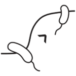
The formation and organization of subcellular domains is critical for numerous cellular processes, even in relatively simple unicellular organisms such as bacteria. Our lab is interested in exploring the factors and mechanisms that generate the subcellular and functional organization of bacterial cells, particularly focusing on development and organization of the cell pole. Cell poles constitute key subcellular domains that are often critical for multiple cellular functions.
We use Vibrio cholerae, a motile Gram-negative rod with unipolar flagellum as model organism. Unlike many other bacteria, the genome of V. cholerae is divided into two circular chromosomes (Yamaichi et al., 1999) that have been shown to possess replicon-specific systems for replication initiation and segregation, and V. cholerae cells can grow very rapidly without mis-segregation of the two chromosomes.
We identified V. cholerae polar anchor protein HubP, which tether multiple proteins involved in motility, chemotaxis, and chromosome segregation thus functioning as cell pole organizer (Yamaichi et al., 2012).
We aim comprehensive understanding of cell pole organization and its development, by using different approaches including comparative proteomics (Altinoglu et al., 2022) and super resolution mircoscopy (Altinoglu et al., 2019).
Selected publications:
Yamaichi, Y., Bruckner, R., Ringgaard, S., Möll, A., Cameron, D.E., Briegel, A., Jensen, G.J., Davis, B.M., Waldor, M.K. (2012) A multidomain hub anchors the chromosome segregation and chemotactic machinery to the bacterial pole. Genes Dev. 26: 2348-2360.
https://doi.org/10.1101/gad.199869.112
Altinoglu, I., Merrifield, C.J., Yamaichi, Y. (2019) Single molecule super-resolution imaging of bacterial cell pole proteins with high-throughput quantitative analysis pipeline. Sci Rep. 9: 6680.
https://doi.org/10.1038/s41598-019-43051-7
Altinoglu, I., Abriat, G., Carreaux, A., Torres-Sánchez, L., Poidevin, M., Krasteva, P.V., Yamaichi, Y. (2022) Analysis of HubP-dependent cell pole protein targeting in Vibrio cholerae uncovers novel motility regulators. PLoS Genet. 18(1): e1009991
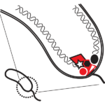
Replication and segregation of bacterial genomes manifest faithful inheritance of the genetic information through many generations. We investigate subcellular organization of the chromosomes as well as ParABS partitioning system-dependent and ParABS-independent chromosome segregation mechanisms.
Selected publications:
Yamaichi, Y., Bruckner, R., Ringgaard, S., Möll, A., Cameron, D.E., Briegel, A., Jensen, G.J., Davis, B.M., Waldor, M.K. (2012) A multidomain hub anchors the chromosome segregation and chemotactic machinery to the bacterial pole. Genes Dev. 26: 2348-2360.
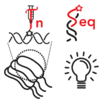
Transposon-insertion sequencing (Tnseq) combines transposon mutagenesis and next generation sequencing (NGS) methodologies to assess essentiality and/or fitness contribution for genes and genetic loci in given growth condition. We used Tnseq to understand transfer capacity of conjugative plasmid (Yamaichi et al., 2015) as well as genetic interactions in V. cholerae.
Our collaborator François-Xavier Barre in I2BC, recently developed Hi-SC2, a new Tnseq-based method to monitor sister-chromatid contacts (Espinosa et al, 2020a, 2020b).
We are interested in novel genome-wide survey technique that allow us to address certain biological questions that were not able to tackle heretofore.
Selected publications:
Yamaichi, Y., Chao, M.C., Sasabe, J., Clark, L., Davis, B.M. Yamamoto, N., Mori, H., Kurokawa, K., Waldor, M.K. (2015) High-resolution genetic analysis of the requirements for horizontal transmission of the ESBL plasmid from Escherichia coli O104:H4. Nucleic Acids Res. 43: 348-360.
https://doi.org/10.1093/nar/gku1262
Yamaichi, Y. and Dörr, T. (2017) Transposon insertion site sequencing for synthetic lethal screening. Methods Mol Biol. 1624: 39-49.
https://doi.org/10.1007/978-1-4939-7098-8_4
Espinosa, E., Yamaichi, Y., Barre, F.-X. (2020b) Protocol for High-throughput analysis of sister-chromatids contacts (Hi-SC2). STAR Protoc. 1: 100202.
DNA modifications that do not change the nucleotide code (G/A/T/C) can affect gene activity. Besides such ‘epigenetic’ feature, DNA modification have other biological implications. In Bacteria, one well-known mechanism is called restriction-modification (R-M) system. In general, restriction enzymes recognize specific nucleotide sequence and digest the double-strand of DNA (as you know, they are exactly the “restriction enzymes” we use for molecular biology). Modification enzymes also recognize the nucleotide sequence, but they modify (methylate) the nucleotide and resulting DNA became tolerant to cognate restriction enzyme. Together, R-M system works as defense mechanism from alien DNA intake.
a) Our model conjugative plasmid pESBL encodes very atypical, orphan DNA methyltransferase (without restriction enzyme) called M.EcoGIX. In the first report M.EcoGIX does not have specific recognition sequence but methylate whatever A residue (Fang et al., 2012). It was later refined to target SAY motif, yet methylate A very often (Fomenkov et al., 2020). More interestingly, M.EcoGIX only methylates one of the two strand of DNA (Fang et al., 2012).
Biologically, we showed evidence that M.EcoGIX counteracts R-M system of the recipient cells, thus facilitates establishment of pESBL in new host cells (Yamaichi et al., 2015).
Further study, with collaboration with New England Biolabs, suggested that M.EcoGIX enzyme activity depends on DNA polymerase I. Expression and action of M.EcoGIX appear to occur in the recipient cell at the very early stage of conjugational transfer where single strand DNA is transferred and converted into double stranded DNA (Fomenkov et al., 2020).
b) Natural host of pESBL (EHEC O104:H4) also encoded other DNA methyltransferases M.EcoGI/II. M.EcoGI/II also showed unspecific methylation activity (Fang et al., 2012). Taking advantage of this unique feature, former colleague in I2BC, Laure Crabbé, developed MAD-ID technique (Sobecki et al., 2018. M.EcoGII is currently available from New England Biolabs with “Enzymes for Innovation” tag.
We are interested in biology and application of M.EcoGIX and other atypical DNA methyltransferases.
Selected publications:
Yamaichi, Y., Chao, M.C., Sasabe, J., Clark, L., Davis, B.M. Yamamoto, N., Mori, H., Kurokawa, K., Waldor, M.K. (2015) High-resolution genetic analysis of the requirements for horizontal transmission of the ESBL plasmid from Escherichia coli O104:H4. Nucleic Acids Res. 43: 348-360.
https://doi.org/10.1093/nar/gku1262
Fomenkov, A., Sun, Z., Murray, I.A., Ruse, C., McClung, C., Yamaichi, Y., Raleigh, E.A., Roberts, R.J. (2020 Plasmid replication-associated single-strand-specific methyltransferases. Nucleic Acids Res. 48: 12858-12873.
Bacteria display wide variety of shapes, and it has been thought that selective forces allowed them to evolve different forms favorable to particular niches. Yet, bacteria share common molecule, peptidoglycan (PG), as the major component of the cell wall. Bacterial cell growth requires insertion of new PG monomer and asymmetric insertion of new PG can define certain cell shape such as curved rod or helical shape. Furthermore, coordinated PG hydrolysis is essential for successful cell division. Therefore, PG has been a major target of antimicrobial agents such as penicillin.
Bacteria also change cell shape at different growth conditions, for example Escherichia coli can change cell shape between exponentially growing phase and stationary phase. Similar to E. coli, V. cholerae cells from overnight cultures (stationary phase) are much smaller than those from exponentially growing culture (log phase). Previously, it has shown that V. cholerae cells ‘remodel’ PG when entering the stationary phase, as environmental D-amino acids are accumulated (like quorum sensing).
We are interested in molecular mechanisms how bacterial cells exhibit such physiological changes at different growth stages.

