CryoEM
Cryo-electron microscopy
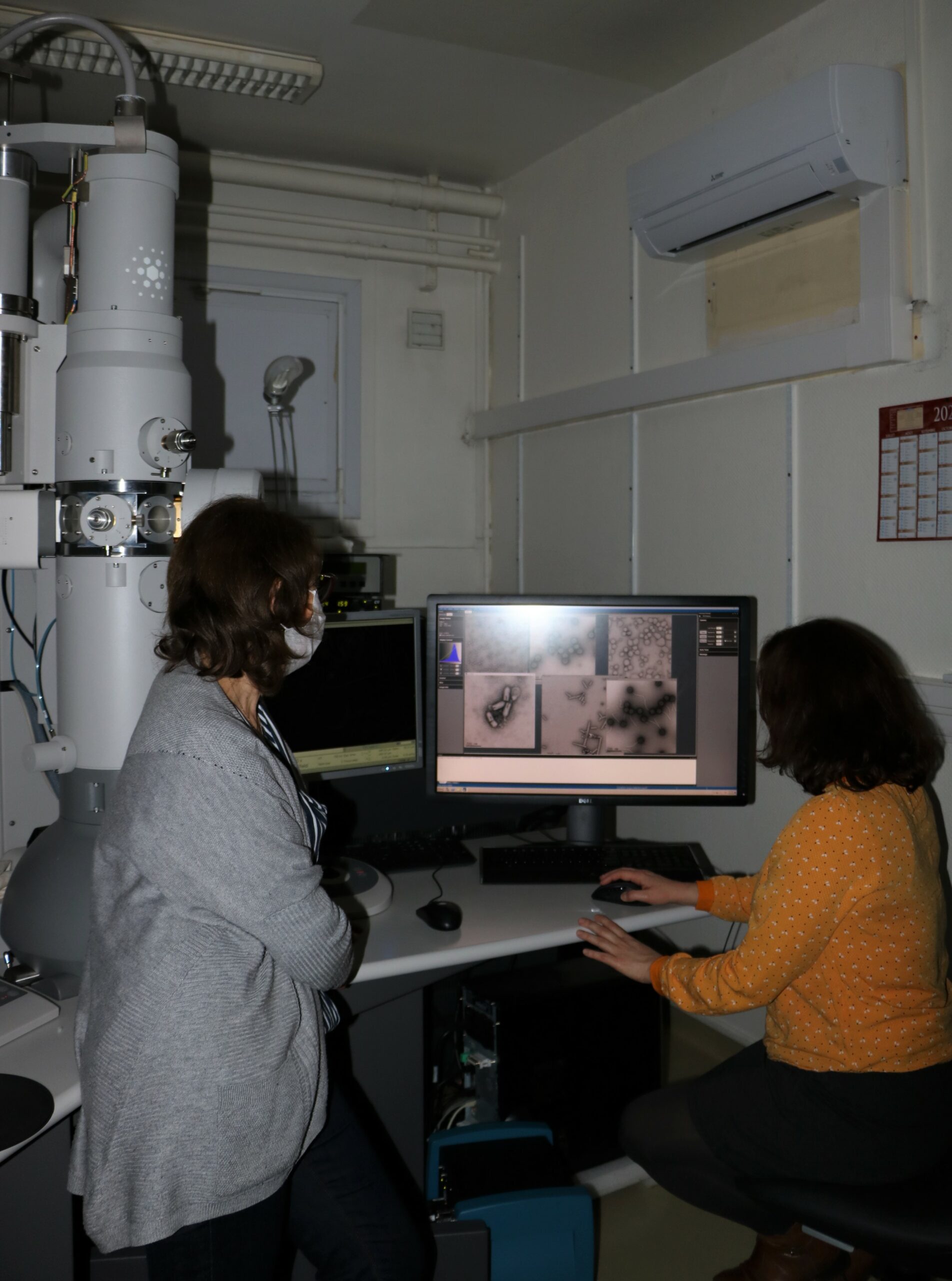
Cryo-electron microscopy visualizes biological objects in their natural aqueous environment.
Cold stage stability and the properties of the camera determine the resolution of the structural data obtained (1-0.25 nm).
KeyTech
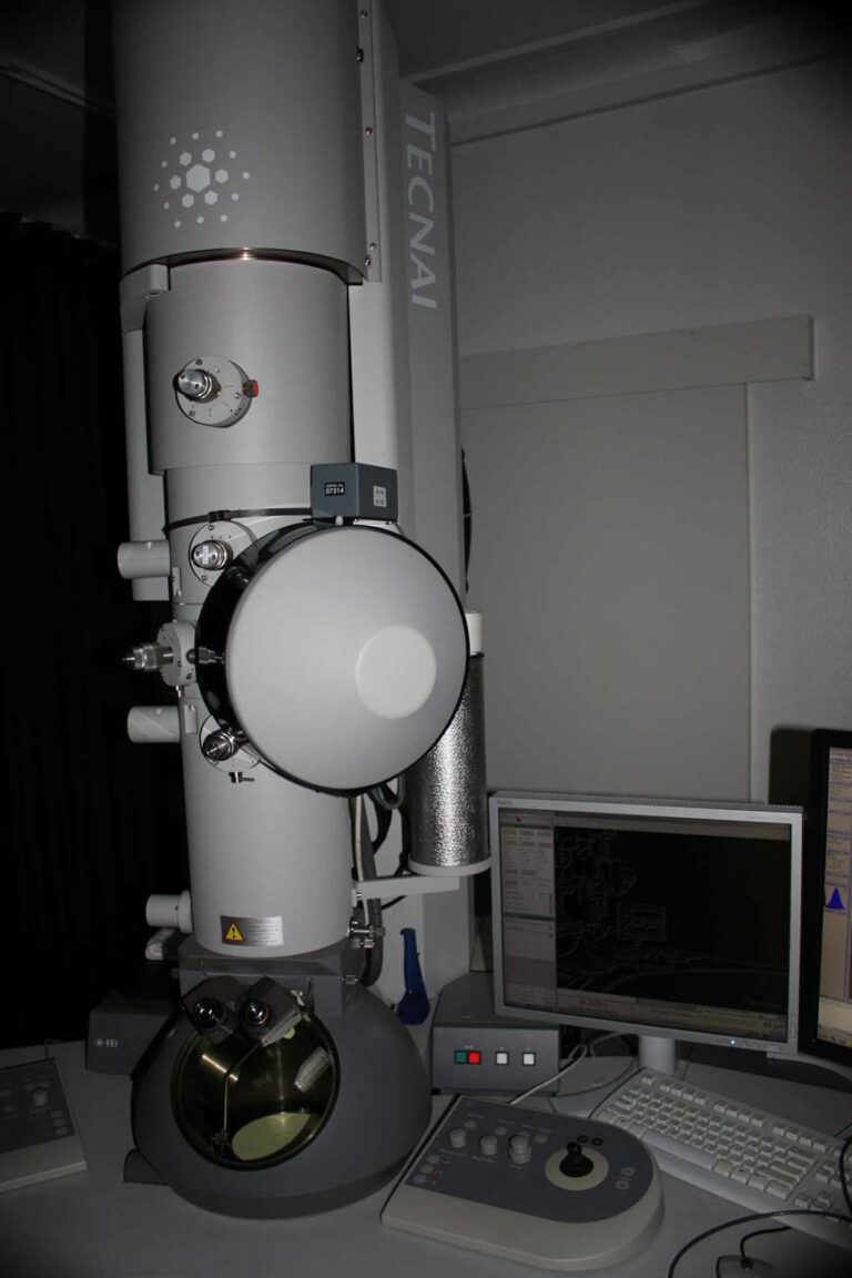
The cryo-electron microscopy facility currently comprises two cryo-electron microscopes 200 kV and 120 kV (Tecnai FEI), each equipped with a direct detection camera (K2 Gatan) and a liquid nitrogen cooled holder (Gatan 626). The 200 kV microscope is equipped with a field emission gun (FEG) and a camera that detects single electrons (Gatan K2 summit); the 120 kV has a LaB6 filament and a basic direct detection camera (Gatan K2 base).
The present FEG 200 kV cryo-electron microscope has a side-entry stage that is less stable than top entry holders. However, the microscope is equipped with a direct detection camera that, due to its high sensitivity, allows the recording of movies composed of frames with short time exposure (0.1s). Centering and averaging of images from the movie can compensate for the holder drift, leading to high signal to noise ratios. The 200 kV cryo-electron microscope of the facility is thus an ideal tool to initiate studies and to obtain an initial model at a resolution better than 1 nm. Such a model will be very useful to determine the atomic structure of the object with state of the art microscopes, for instance the 300kV available at Institut Pasteur or Grenoble.

200kV cryo-electron microscope equipped with a FEG, a liquid-nitrogen

Access to the microscopes is provided in three stages:
- limited service,
- use
- collaboration.
The facility engineers will first test by both conventional and cryo-electron microscopy the feasibility of the potential projects. Those projects which show potential can be continued on higher end microscopes, possibly with the help of the facility engineers for further grid screening and/or data collection.
The facility has the necessary computing capabilities for carrying out image processing and 3D reconstruction to atomic resolution.
Engineers can advise, assist and/or collaborate at all steps on a case by case basis, depending on user experience and sample quality. The facility has experience in both single particle analysis and helical reconstruction.
The microscopes are installed in a BSL2 laboratory, and so projects requiring this security environment can be considered with the agreement of the Scientific committee of the department.

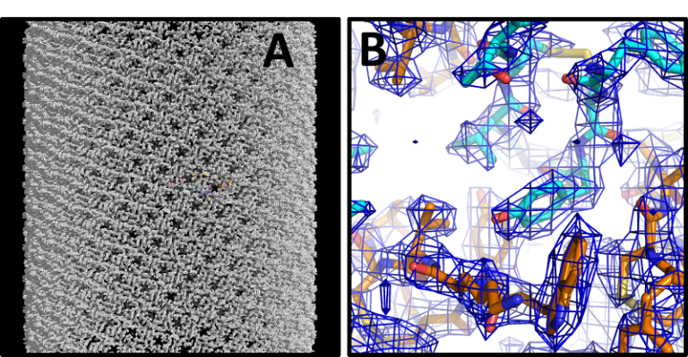
Lanreotide is a somatostatin hormone analog used against acromegaly and for certain forms of neuroendocrine cancers. Its sustained-release formulation consists of only water and peptide that self-assembles into 250 Å diameter nanotubes (A) reconstructed here to 2.5 Å resolution (B) (Pieri et al., PNAS 2022)
team

Stéphane BRESSANELLI
Head of Facility
Research Director

Aurélie ALBERTINI
Deputy Head
Researcher

Ana Andreea ARTENI
Technical Manager
Research Engineer
Malika OULD ALI

Engineer
Laura PIERI

